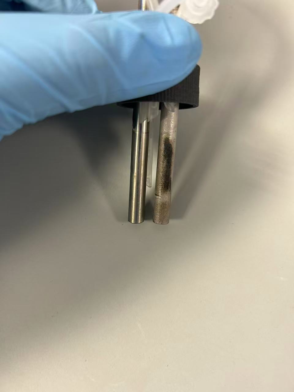
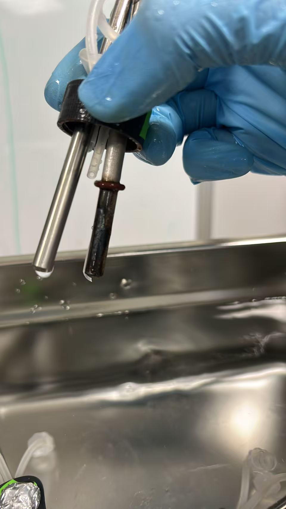
Here’s a rather pretty image of the discolouration from the anode that @Bingqiao returned to me:
At the bottom, above the shadow, we see the brown discolouration that we found on the cathode-facing side of the anode. Above this you see a transition to rainbow discolouration. At the very top you can see the natural platinum colour.
My initial fears were that this was caused by loss of platinum, as titanium is well known for taking on rainbow oxidation patterns. There are two things that give me hope that this isn’t the case:
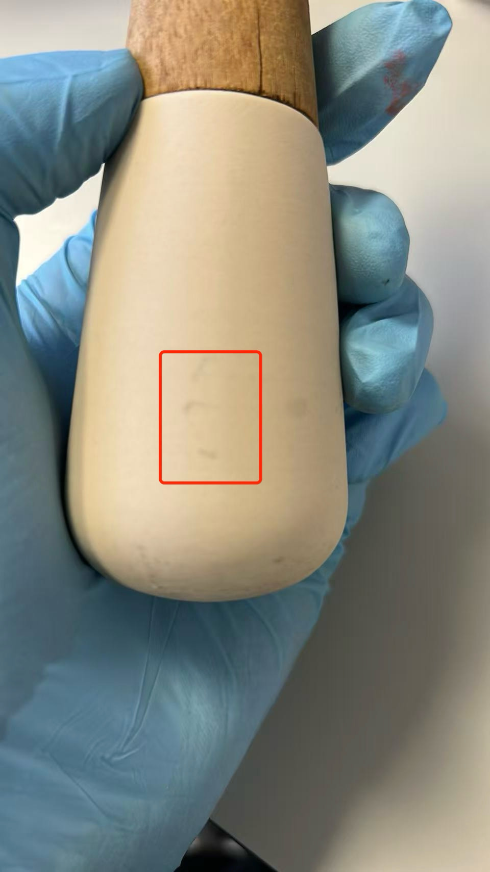
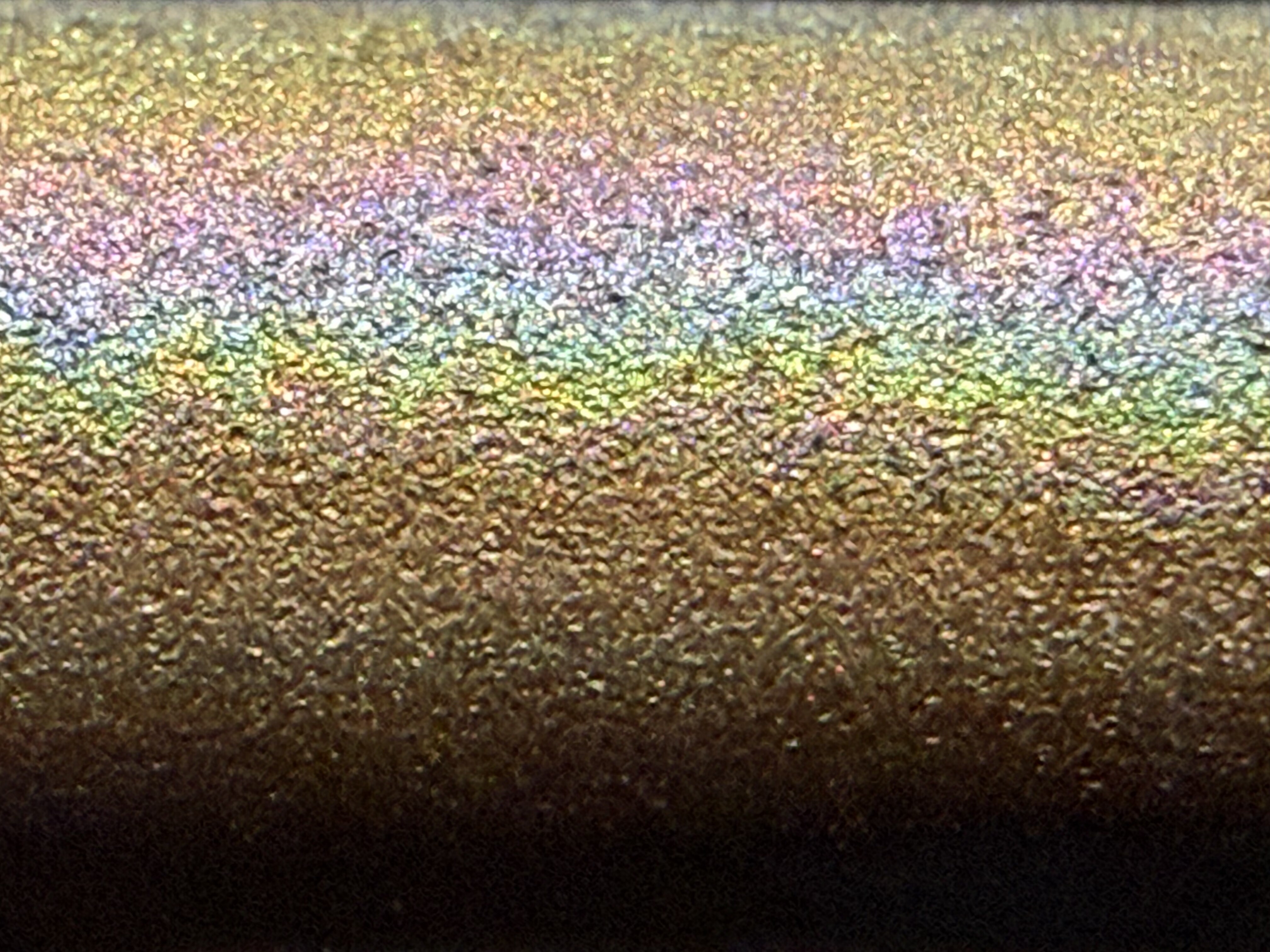
Firstly, in this image of the top of the electrode:
The left of shows the un-plated titanium, with plated on the right. The discoloured section appears to have the same platinum plated texture. It could be that the platinum is porous and the titanium is oxidising beneath it, but to me it looks like the rainbow is relatively continuous and on the surface of the textured platinum.
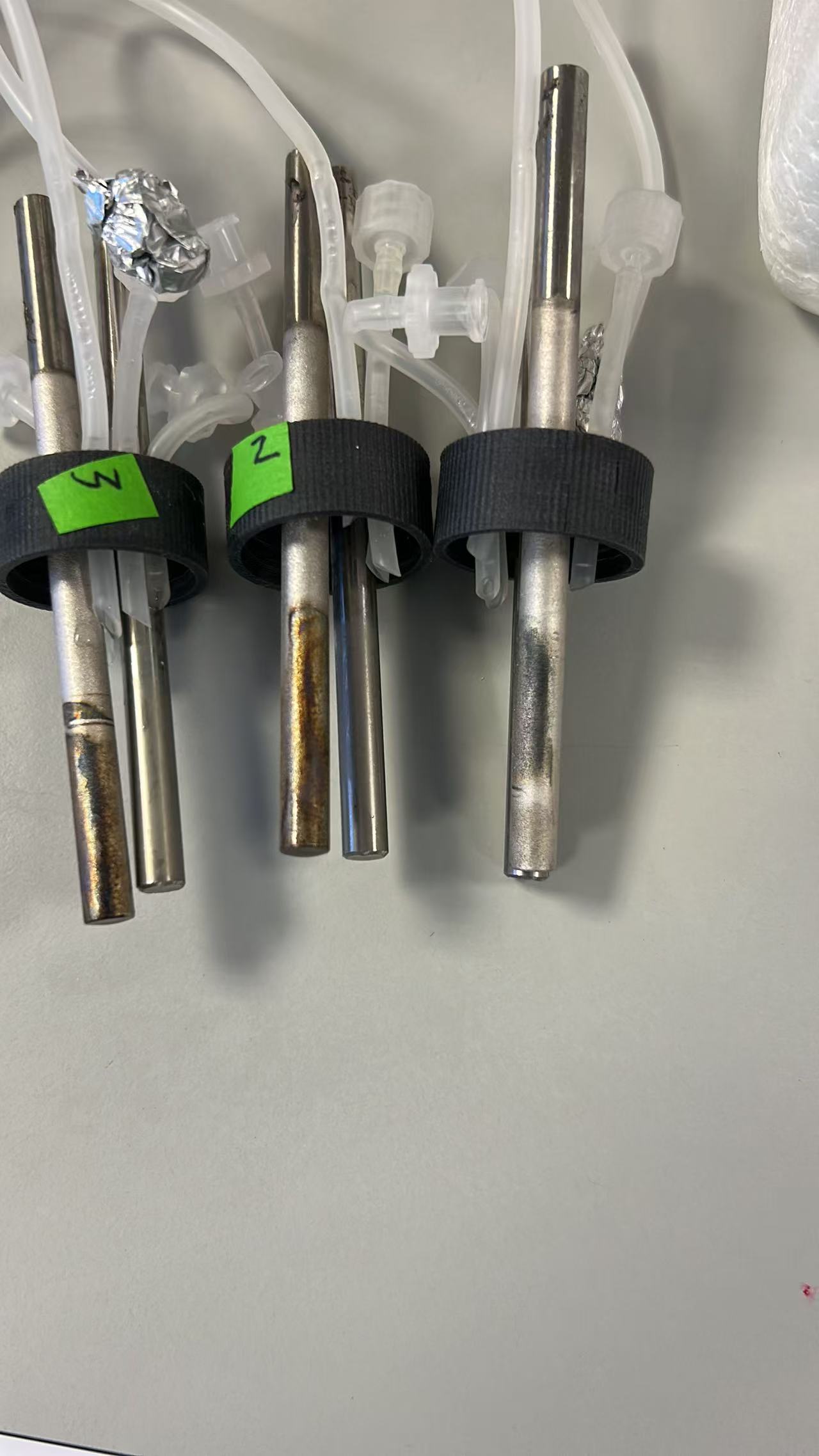
The second is thanks to @gerrit stepping in to help. The depicted electrode arrived the day I went on leave, and I’ve been off work since with man-flu that I went down with on my first day back. I’ve been using PHREEQC to determine the theoretical conductivity of the solution we were working with - unfortunately I wasn’t taking notes during the training sessions so don’t have the measured conductivity or even temperature.
My plan was to run electrolysis at the same conductivity but with a single salt. Gerrit gave this a go while I was still running calculations. I understand he used excess sodium bicarbonate and ran at 10% intensity (measuring 13 mA and ~4.9V) for over 20h without seeing any discolouration.
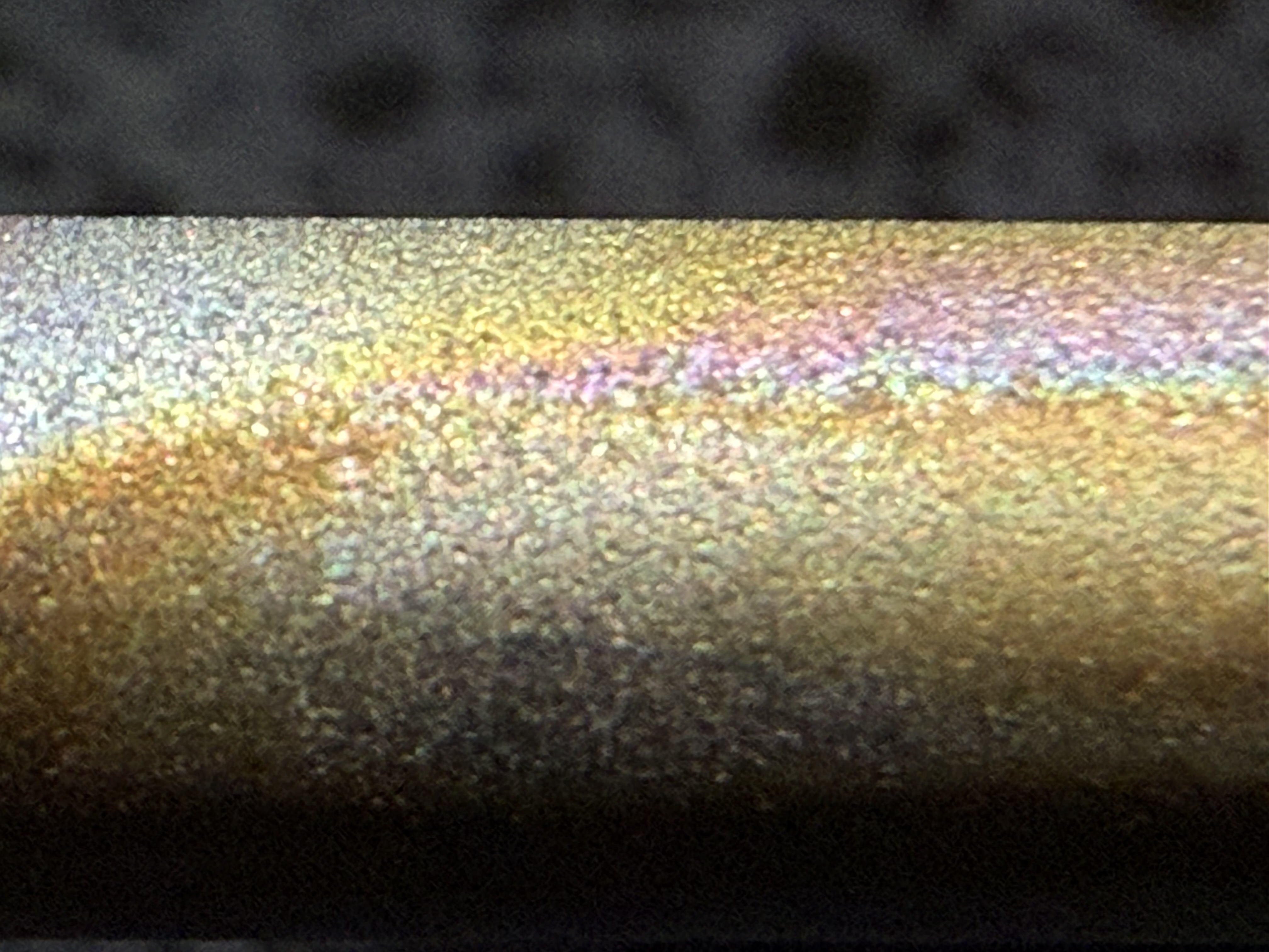
These results combined made me wonder if what we are seeing is ferric (phosphate?) deposition at visible thicknesses in the brown area and wavelength thicknesses in the rainbow area.
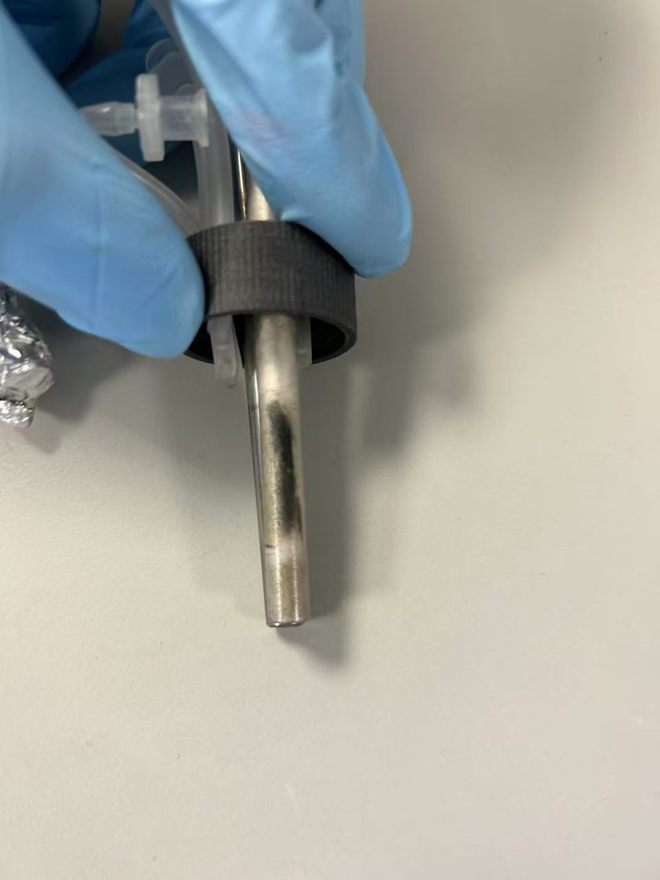
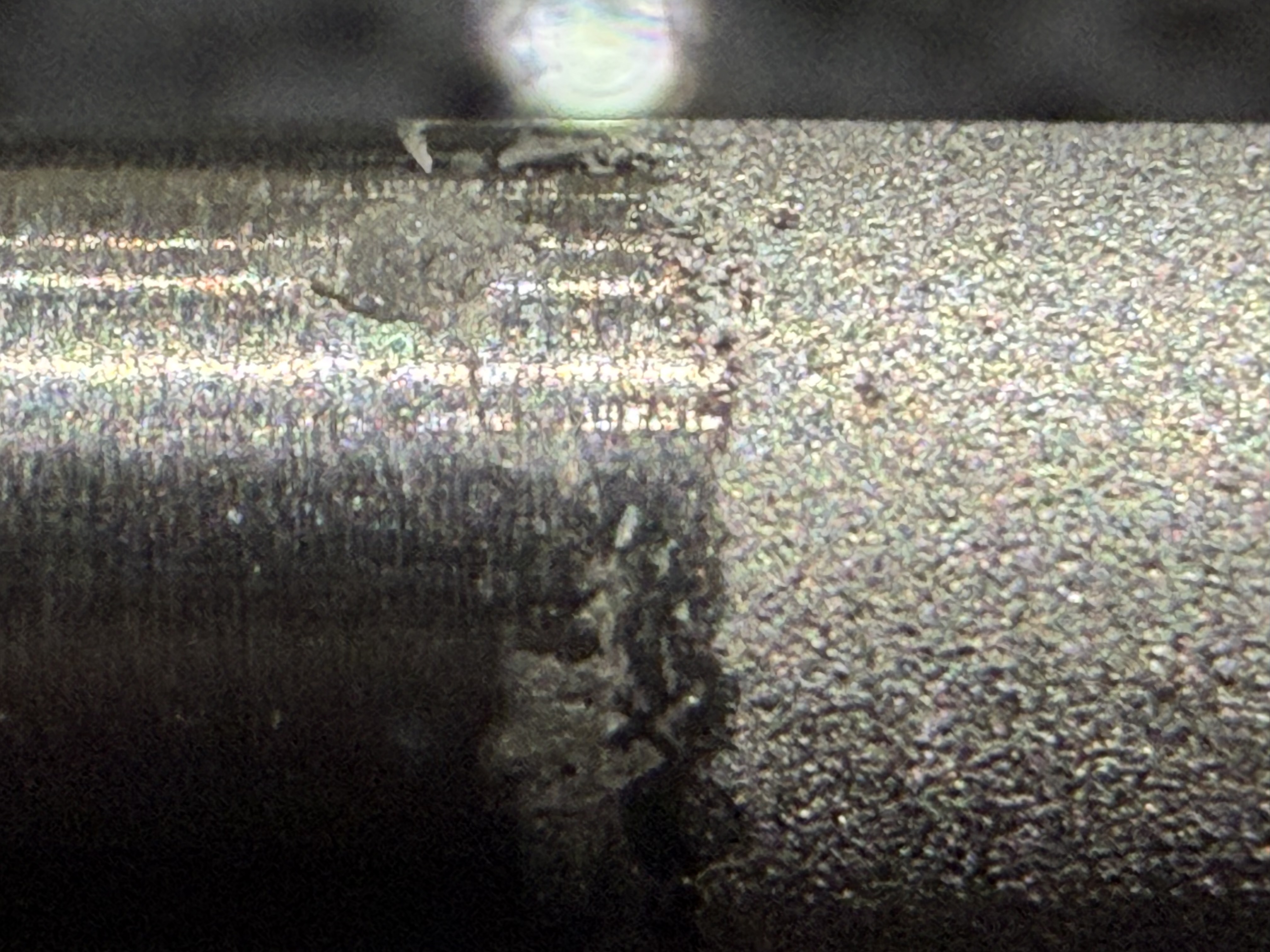
I haven’t been well enough to go out for strong acid, but swabbing the second quarter from the left in this image with distilled barley malt vinegar:
…visibly transferred some brown to the swab. That suggests deposition. I’m a little concerned that the rainbow wouldn’t lift there, but hope that it will with a strong acid.
So, I’m hopeful that we are not irreparably impacting the platinum plating, but if this is ferric phosphate deposition, it will reduce our effective anode area and increase required overpotential. Chris French mentioned switching away from phosphate previously. This may be another reason to try that.